What are the Basic Principles using in ECG?
THE BASIC PRINCIPLES OF ELECTROCARDIOGRAPHY
Electrocardiography (ECG) has come a long way since Willem Einthoven invented the first practical
electrocardiograph in 1903. The first machines were very large and required three feet to be submerged in jars filled with salt water to obtain an EKG. The EKG records the electrical activity of the heart. The heart's electrical activity usually begins at the sinus node (SA) in the upper right corner of the right atrium and progresses downward toward the ventricle. Transmission from the SA node to the next relay station, called the atrioventricular (AV) node, in the lower right atrium occurs via the internodal pathway, a special conducting tissue in the right atrium.
One of the internodal pathways extends into the left atrium, through which conduction simultaneously passes into the left atrium. At the AV node, there is a short delay in the conduction of the impulse, after which it reaches the bundle of His to pass forward to the ventricles. Its bundle has two divisions - the right and left bundle branches for the two ventricles. The left branch has two anterior and posterior, sometimes a third middle branch. This division is called Da Nang. ECG is often absorbed by an electrode attached to another part of the body surface.
If you take it as an electrode stored in the center of the registration, it will be carried out in a study of invasive electrical physiology in an electrical physiological laboratory. Typically, an EKG uses 4 leg electrodes on each of its 4 legs, of which the electrode on the right leg is considered electrically neutral and the remaining 3 are active electrodes. In addition, six electrodes are placed in specially designated areas of the chest cavity to obtain images of the chest cavity. Although only 10 electrodes are used to record a typical ECG, the most common recording is the 12-lead, as these electrodes can record different combinations of leads. In special cases, additional electrodes can be used to increase the number of wires. Waves, segments, and intervals in the ECG
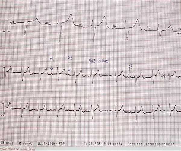
ECG waves and segments
The waves seen in a normal EKG are called the P wave, QRS complex, T wave, and sometimes U wave. Other less common waves are delta waves and epsilon waves. The P wave represents the depolarization of the atria, and the QRS complex represents the depolarization of the ventricles. T waves are associated with ventricular repolarization. The PR segment lies between the P wave and the QRS complex and the ST segment lies between the QRS complex and the T wave.
The TP segment is located between the T wave and the next P wave. It is considered the true baseline for the ECG. Intervals contain one or more waves and segments. The PR interval includes the P wave and the PR segment and is measured from the beginning of the P wave to the beginning of the QRS complex. The QT interval consists of the QRS complex, the ST segment, and the T wave. It is measured from the beginning of the QRS complex to the end of the T wave.
The PP interval is measured from the beginning of one P wave to the beginning of the next. The RR interval is measured from the beginning of one QRS complex to the beginning of the next, or, for simplicity, the interval between the peaks of two consecutive R waves. In certain pathological conditions, delta waves appear at the beginning of the QRS complex and epsilon waves appear at the ends of the QRS complex. In hypokalemia, there is a prominent U wave after the T wave.
The components of the QRS complex
The initial sound deflection that is part of the QRS complex is called the Q wave. The initial positive deflection is called the R wave. The second negative deflection or negative deflection after the R wave is called the S wave. The second positive deflection is called the R' wave. A negative deviation after R' is called S'. Waves with amplitudes less than 5 mm may be written in lowercase, so rare represents r minor followed by normal S waves and r minor. rSR' indicates a good amplitude of the ending R' wave.
A little about U waves
In the ECG, the U wave appears after the T wave and usually occurs in the middle precordial lead. In hypokalemia, the T wave is flattened and the U wave is pronounced. The main conditions associated with U waves include systemic hypertension, aortic and mitral valve insufficiency, and coronary artery disease.
The theory about the origin of U waves
One theory for the origin of the U wave is that it is due to the repolarization of the Purkinje fibers. Another possibility could be posterior dislocation due to mechanical forces on the ventricular wall. A third hypothesis suggests that this is due to prolonged repolarization of central myocardial M cells. Another hypothesis is delayed repolarization of the papillary muscles. Neither of these theories is widely accepted.
Significance of a negative U wave
An inverted U wave usually has the same meaning as an inverted T wave. A negative U wave is considered highly specific for heart disease and is associated with other abnormalities on the ECG in 90% of cases. Discordant U-wave inversion (including vertical T-wave inversion) is also considered to indicate myocardial ischemia. An interesting case of U wave caused by right ventricular compression of a reconstructed gavage after surgery for esophageal cancer was reported. Persistent U-wave inversion has been observed in cases after anaerobic brain injury.
Exercise-induced U-wave inversion
Exercise-induced U-wave inversion of the anterior lead is in several cases associated with stenosis of the proximal left anterior descending coronary artery. you in turn waving your hand
U-wave shifts with pulse shifts have been described in left ventricular failure.

An inquisitive individual with a great interest in the subjectivity of human experiences, behavior, and the complexity of the human mind. Enthusiased to learn, volunteer, and participate. Always driven by the motive to make a difference in the sphere of mental health - and normalize seeking help through a sensitive and empathetic approach